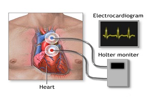
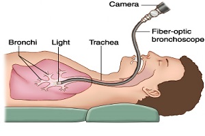
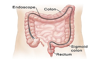
2017-09-23 02:13:19 18 It is a portable EKG that monitors electric activity of heart for one or two days. It may be advised if the doctor suspects an abnormal heartbeat, or palpitations or not enough blood flow to your heart. This works similar to ECG with electrode patches placed on the skin. But in this procedure, you are allowed to go home and continue with all normal activities in the house except taking a shower. This is advised only if symptoms occur every now and then. Sometimes this device may be required to be worn for weeks or even months to record unusual activity in the heart. This can be recorded by pushing a button every time the symptom arises. The device will record the electric activity during that time. The reading is sent to your doctor who will analyze it and judge the gravity of the situation. 2017-09-23 02:13:19 19 Bronchoscopy is a procedure, where a thin viewing tube is inserted through your mouth. This viewing apparatus is called a Bronchoscope. This procedure is done to examine your throat, larynx, trachea, and the full airways. This is an outpatient procedure. This procedure is performed both for diagnostic and therapeutic purposes. Generally, this procedure is advised if the doctor suspects you to have an abnormal chest X-ray or a scan that shows infection or tumor. In some cases a collapsed lung might also be a reason to perform the test. Sometimes this procedure also helps to remove food particles that are stuck by mistake in the airways. This test is also performed to retrieve sample tissue for further examination (biopsy). This test might take from 15 to 45 minutes and the results of this test take from 1 to 4 days. 2017-09-23 02:13:19 20 Lower GI endoscopy allows your doctor to view your lower gastrointestinal (GI) tract. Your entire colon and rectum can be examined (colonoscopy). or just the rectum and sigmoid colon can be examined (sigmoidoscopy). It is performed as an outpatient procedure under sedation .The most common indication for colonoscopy or sigmoidoscopy is bleeding per rectum or blood in stools, altered bowel habits ,constipation or your doctor suspects a tumor or cancer of your lower GI tract. It can diagnose the cause for the bleeding and sometimes can treat it in the same sitting without the need for surgery. It also helps to take biopsies of growth or tumor and can remove polyps of rectum and large intestines.
There are some instructions to be followed before colonoscopy. For a colonoscopy, you may be told not to to drink only clear liquids for 1 day before the procedure. You are instructed to take some laxative preparation before the procedure.
Colonoscopy can take 30 minutes or longer. Sigmoidoscopy often takes about 20 minutes. The length of the procedure depends on how clean your intestines are, the reason for the procedure, and what treatments must be done.
During colonoscopy the doctor uses a colonoscope, a long, flexible, tubular instrument about 1/2-inch in diameter that transmits an image of the lining of the colon so the doctor can examine it for any abnormalities. The colonoscope is inserted through the rectum and advanced to the other end of the large intestine .The endoscope carries images of your colon to a video screen. Prints of the images may be taken as a record of your exam.
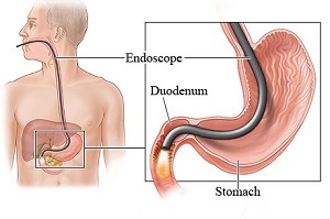
2017-09-23 02:13:19 21 An upper gastrointestinal (UGI) endoscopy is a procedure that allows your doctor to look at the inside lining of your esophagus, stomach and first part of small intestine called duodenum. A thin, flexible viewing tool called an endoscope (scope) is used. The tip of the scope is inserted through your mouth and then gently moved down your throat into the esophagus, stomach and duodenum (upper gastrointestinal tract). This procedure is sometimes called esophagogastroduodenoscopy (EGD).
It is usually done in patients to find the cause in patients who experience vomiting, abdominal bloat, unexplained weight loss,difficulty in swallowing, gastroesophageal reflux disease or if the doctor suspects any tumour of the esophagus or stomach based on your symptoms. It is done as an out patient procedure. Sometimes endoscopy is indicated on Emergency basis if to check for an injury to the esophagus.(For example, this may be done if the person has swallowed poison.) or to remove foreign objects that have been swallowed or to find and treat the cause for vomiting blood.
Using the scope, your doctor can look for ulcers, inflammation (Esophagitis,Gastritis), narrowing of esophagus (stricture),enlarged or swollen veins in the esophagus or stomach(These veins are called varices) , tumors, cancers, infection, hiatus hernia, gerd or bleeding. It can collect tissue samples (biopsy), remove polyps, and treat bleeding through the scope.
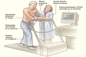
2017-09-23 02:13:19 22 Tread Mill Exercise testing is a cardiovascular stress test that uses treadmill bicycle exercise with electrocardiography (ECG) and blood pressure monitoring.Exercise stress testing, which is now widely available at a relatively low cost, is currently used most frequently to estimate prognosis and determine functional capacity, to assess the probability and extent of coronary disease, and to assess the effects of therapy.
Cardiovascular exercise stress testing in conjunction with ECG has been established as one of the focal points in the diagnosis and prognosis of cardiovascular disease, specifically coronary artery disease (CAD).
2017-09-23 02:13:19 23 A Doppler ultrasound uses reflected sound waves to see how much blood flows through a blood vessel. The doctor can assess the blood flow through major arteries, veins and recognize any changes like blocked or reduced flow of blood. This problem could cause stroke.
There are 3 basic types of Doppler ultrasound-
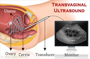
2017-09-23 02:13:19 24 An Ultrasound machine produces high frequency sound waves which passes through the human body and gets reflected back. The reflected back sound waves are integrated to produce an image of the body parts on the monitor. These images are analyzed by the doctor to identify the problems of Uterus, Ovaries, Fallopian tubes, Cervix and vagina.
Transvaginal ultrasound is introducing sound waves through the vagina. The machine is connected to a transducer or a wand which is introduced into the vagina for imaging.
When is transvaginal sonography performed?
Gynecological indications-
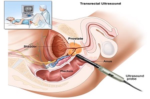
2017-09-23 02:13:19 25 A transrectal ultrasound (TRUS) is a procedure which utilizes sound waves to create an image of the prostate gland and the surrounding tissue. Generally the procedure requires the insertion of an ultrasound probe into the patient’s rectum. The probe then both sends and receives sound waves through the rectal wall into the prostate gland which is situated directly in front of the rectum.
A transrectal ultrasound is done for a number of reasons which include:
2017-09-23 02:13:19 26 An echocardiogram (echo) is a test that uses high frequency sound waves (ultrasound) to take pictures of your heart. The test is also called echocardiography or diagnostic cardiac ultrasound.
An echo uses sound waves to create pictures of your heart’s chambers, valves, walls and the blood vessels (aorta, arteries, veins) attached to your heart.A probe called a transducer is passed over your chest. The probe produces sound waves that bounce off your heart and “echo” back to the probe. These waves are changed into pictures viewed on a video monitor.
Your doctor may use an echo test to look at your heart’s structure and check how well your heart functions. The test helps your doctor find out:
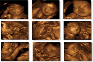
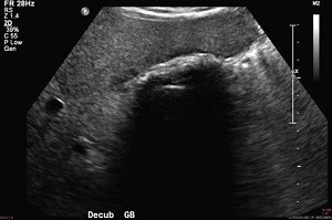
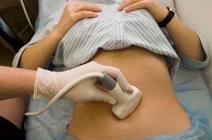
2017-09-23 02:13:19 27 An ultrasound scan is a medical test that uses high-frequency sound waves to capture live images from the inside of your body. It’s also known as sonography. Unlike other imaging techniques, ultrasound uses no radiation. For this reason, it’s the preferred method for viewing a developing fetus during pregnancy. Your doctor may order an ultrasound if you’re having pain, swelling, or other symptoms that require an internal view of your organs.
An ultrasound can provide a view of the urinary bladder, gallbladder, appendix,kidneys ,liver, spleen, pancreas, uterus, ovaries, thyroid, testicles and blood vessels.It is also a helpful way to guide surgeons’ movements during certain medical procedures, such as biopsies. It is the diagnostic tool of choice to diagnose gall bladder stones.
Your doctor may tell you to fast for eight to 12 hours before your ultrasound, especially if your abdomen is being examined. Undigested food can block the sound waves, making it difficult for the sonologist to get a clear picture.For an examination of the gallbladder, liver, pancreas, or spleen, you may be told to eat a fat-free meal the evening before your test and then to fast until the procedure. However, you can continue to drink water and take any medications as instructed. For other examinations, you may be asked to drink a lot of water and to hold your urine so that your bladder is full and better visualized.
If your doctor is able to make a diagnosis of your condition based on your ultrasound, they may begin your treatment immediately. If anything abnormal turn up on the ultrasound or it becomes difficult to diagnose on ultrasound or is inconclusive, you may need to undergo other diagnostic techniques, such as a CT scan, MRI, or a biopsy sample of tissue depending on the area examined.
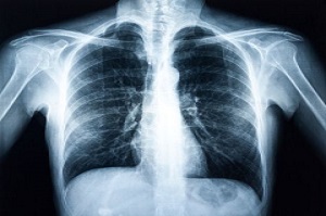
2017-09-23 02:13:19 28 X-ray is a form of electromagnetic radiation. It is similar to visible light, but with a higher energy and they can pass through many objects. It can help the doctor view the inside of your body without having to make any incisions. X-rays can help diagnose and treat many medical conditions. Different types of X-rays are used for different purposes. It can be used on different parts of the body eg. For bone cancer, breast tumors, enlarged heart, blocked blood vessels, conditions affecting your lungs, digestive problems, fractures, infections, osteoporosis, arthritis, tooth decay, retrieve swallowed items.
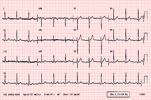
2017-09-23 02:13:19 30 ECG is also called electrocardiogram. It is a test that records the electrical activity of the heart through small electrode patches placed on the skin of your chest, arms, and legs by a technician. The ECG is generally used to check the heart for any irregular blood flow, or to diagnose a heart attack, or anything abnormal with the heart like thickened muscles etc. During the test, the computer creates a picture on a graph sheet created by the electrical impulses that travel through the heart. The whole preparation and the actual test might take about 15 minutes. This test result is kept safe to be compared later with fresh reports as the treatment proceeds.
Why is Doppler done?
How to prepare for Doppler?
How is it done?
Any risks associated with Doppler?
What are the preparations for transvaginal ultrasound?
What happens during transvaginal ultrasound?
What are the risks associated with transvaginal ultrasound?
When do we get the reports?

